
Image of the Month - June 2020
(Photo credit: Stefanie Palmer C.R.A)
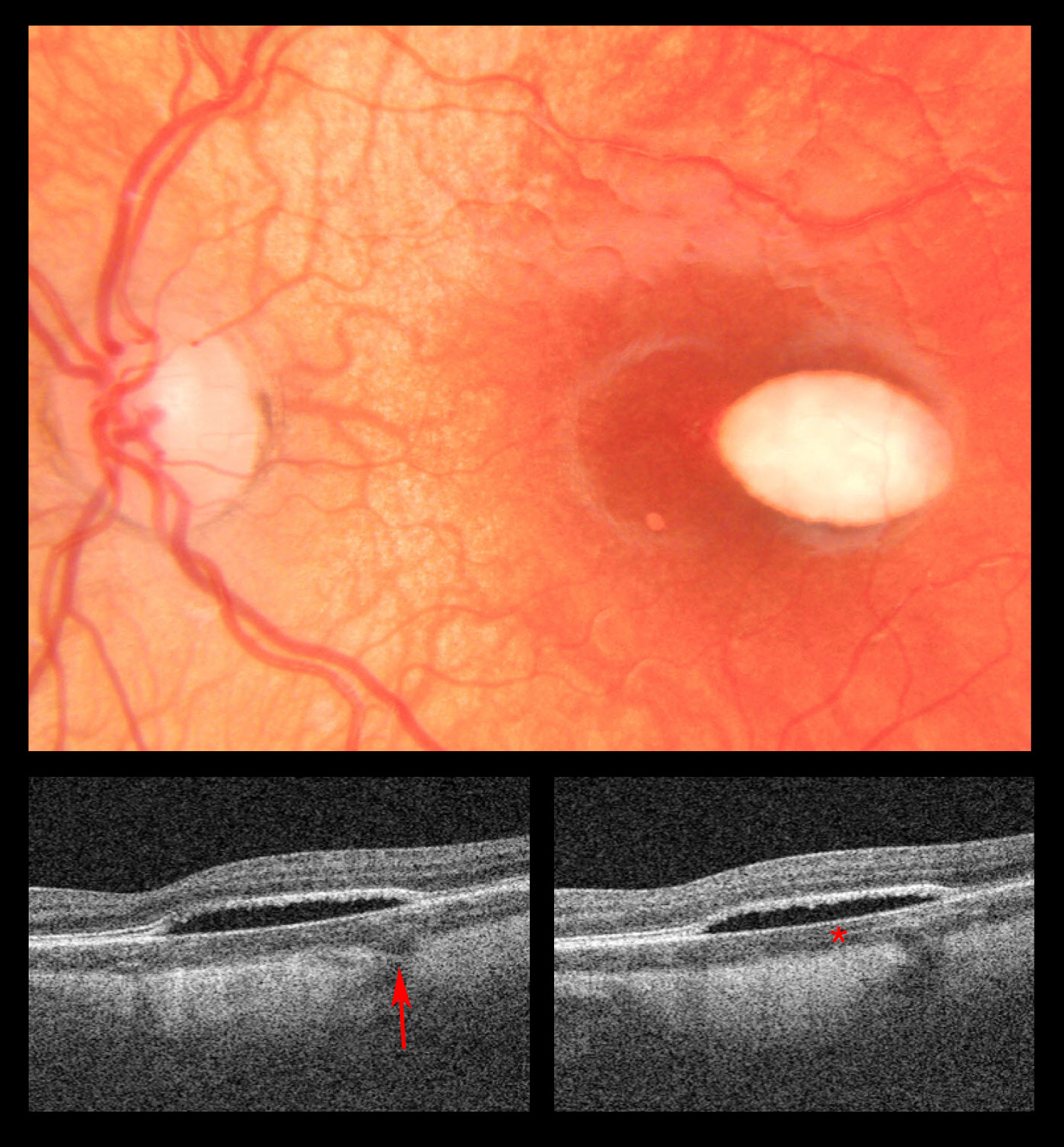
This multi-modal image demonstrates the left eye of a 2 year old patient with a hypopigmented lesion. The fundus photo shows an oval shaped flat hypopigmented lesion in the temporal macula as well as irregular perifoveal reflex and focal RPE atrophy. The corresponding swept-source OCT highlights outer retinal cavitation as well as an abnormality within the choroid layer. There is a focal choroidal excavation posteriorly (arrow) as well as hyperreflectivity in the anterior choriocapillaris region (asterisk). Due to the classic appearance on fundus examination, this lesion most likely resembles torpedo maculopathy. Two distinct categories of torpedo maculopathy have been described in the literature: Type 1 shows attenuation of the outer retina and type 2 shows an outer retinal cavitation. The case presented here may be a variant of type 2 torpedo maculopathy. Torpedo maculopathy is thought to be a congenital condition that remains stable over years without significant impact on vision. Only rare cases of choroidal neovascularization have been reported. The exact etiology of torpedo maculopathy is unknown, and multiple imaging features have been described. We here see abnormalities in the choroid layer in addition to the typical subretinal cleft. This case also highlights the ability for imaging in the pediatric population, which often can be an important adjunct to the clinical exam.