
Anatomy of the Eye
The Outer Eye
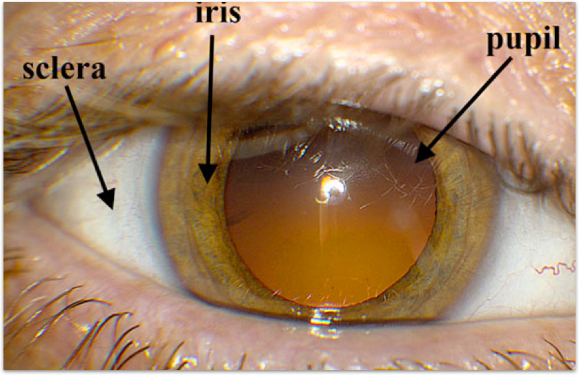
When looking at the outside of the eye, several structures are readily available for viewing. The white part of the eye is a tough outer layer called the sclera. The sclera protects the inside of the eye and helps the eye keep its structure. The colored part of the eye is called the iris. This portion of the eye is what controls the amount of light that is let into the inner eye. It is actually a circular muscle that contracts or relaxes depending on the amount of light it senses. The color of the iris is due to various pigmented cells incorporated into the iris. In the center of the iris is where we find the pupil. The pupil normally looks dark because of light-absorbing pigments in the interior portions of the eye.
The Eye in Cross Section
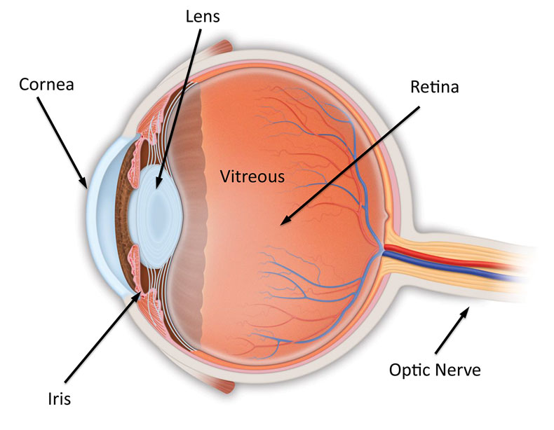
When we look at a cross-section of the eye, we can see the interior structures of the eye. The transparent portion at the very front of the eye is called the cornea. The cornea is a thin layer of cells that allows light to pass through the front of the eye. The lens of the eye sits behind the iris. The lens focuses the light that comes through the cornea onto the back of the eye. Once the light has traveled through the lens, it travels through the transparent vitreous. The vitreous is a water-based viscous jelly-like substance that fills the middle of the eye. It is transparent and also helps to keep the structure of the eye. The retina is a thin layer of neural cells that sits on the back of the eye. The retina is responsible for transmitting the information to the brain through the optic nerve.
The Vitreous
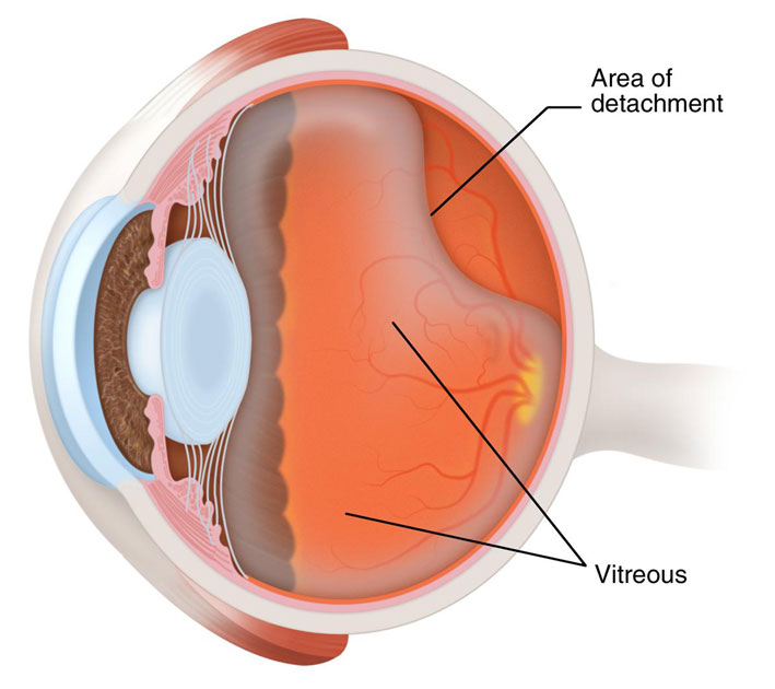
The vitreous is composed of 99% water combined with salts, sugars, and a network of collagen fibers. The other 1% of the vitreous is composed of hyaluronic acid which increases the viscosity of the vitreous to 2 to 4 times that of pure water. The vitreous fills the inside of the eye but does not come in contact with the retina except in three places: the macula (the area of the retina responsible for central vision), the optic nerve disk (where the optic nerve attaches to the eye) and the anterior border of the retina (the border closest to the lens). There are no blood vessels in the vitreous and very few cells. Unlike the fluid in the anterior chamber which is continuously replenished, the vitreous does not turn over. As we age, the vitreous becomes more liquid and degenerates.
The Retina
The retina is a specialized layer of neural tissue that coats the back of the eye. It is an outgrowth of the brain and therefore can be classified as a member of the central nervous system. Although, the retina can easily be compared to the film inside a camera, that analogy is misleading because the retina is very complex. It has many blood vessels that supply nutrients and oxygen to the retinal cells.
The retina is composed of many specialized neural cells. The first layer of the retina to receive visual information is the specialized photoreceptor layer, which is composed of rods (cells responsible for night vision) and cones (the cells responsible for color vision). When excited, the photoreceptors pass their signals along to neighboring neurons in the retina that help to begin processing the visual information and in turn pass their signals on to the brain through the optic nerve.
The Macula
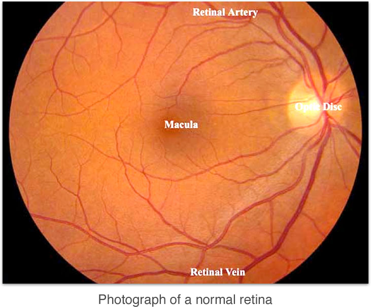
A special part of the retina called the macula is responsible for our central vision. The center of the macula is called the fovea. This area has the greatest concentration of cones and is responsible for most of our fine detailed vision. There are no rods or blood vessels contained in the fovea. We automatically move our eye so that light focuses on the fovea when we want to look at something because it is the area of highest visual acuity. Therefore, the fovea is always responsible for our central vision when we are looking directly at something.
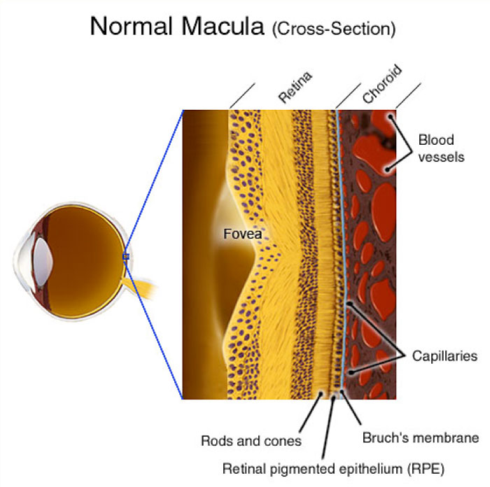
RPE and the Choroid
The retina attaches to the underlying tissue via the retinal pigment epithelium (RPE). The RPE is a highly specialized layer of pigmented cells that play a role in the maintenance of visual function. The highest concentration of pigment in the RPE is found in the peripheral retina and the lowest concentration is in the macula. The RPE acts as an anchor for the retina as well as the blood-retinal barrier. It is involved in metabolism of certain compounds of the retina and the melanin (pigment) in the RPE works to help reduce scattering of light and blocks light absorption through the sclera. Attached to the opposite side of the RPE is a large vascular bed called the choroid. The circulation in the choroid has one of the highest blood flow rates in the entire body per tissue gram. More than 70% of all the blood in the eye is found in the part of the choroid nearest the retina.