
Optical Coherence Tomography
What is Optical Coherence Tomography?
Optical Coherence Tomography (OCT) is a diagnostic imaging device that provides direct high resolution cross-sectional images of the retina. OCT uses a low strength laser to create a 2 or 3-dimensional reconstruction of the retina.
What is the procedure like?
The procedure to obtain an OCT scan of the retina is very quick, safe and painless. To obtain the high-resolution scan of the retina, simply place your chin on the chinrest in front of the machine, look straight ahead at the green star-like crosshair, and keep your eye open for a few seconds without blinking. The entire procedure takes less than about one minute per eye.
How is the OCT image used?
OCT is very useful imaging technique for diagnosing disease. It is especially helpful in evaluating disease of the macula such as: Macular Holes, Vitreomacular Traction Syndrome, Age-related Macular Degeneration, Diabetic Retinopathy, Cystoid Macular Edema, and Central Serous Chorioretinopathy. OCT can help your retinal surgeon to monitor your retinal disease following treatment. Since the images are obtained and processed almost instantaneously, patients are able to view the scans often times together with their physician which greatly helps with their understanding of what is happening inside of their eyes.

Figure 1. Normal OCT.
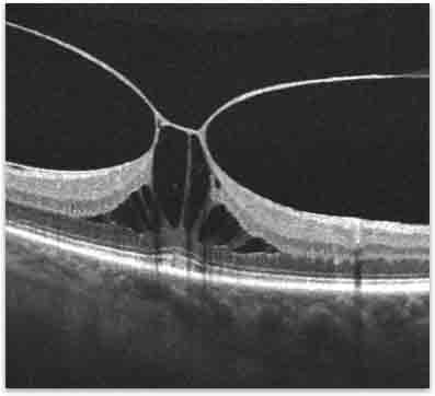
Figure 2. OCT of vitreomacular traction.