
Retinal Tear
What is a Retinal Tear?
The retina is the layer of specialized nerve tissue lining the back of the eye that allows you to see. The inside of the eye is filled with a gel-like substance called the vitreous (figure 1). A retinal tear occurs when there is an abnormal attachment between the vitreous gel and the retina (figure 2). In this case the vitreous gel can tug on the retina causing it to tear. If there are retinal blood vessels at the location of the retinal tear, bleeding will occur inside the eye.
What causes a Retinal Tear and who is at risk?
Retinal tears can develop at any age, but tend to occur more commonly in the elderly. Tears typically occur when the vitreous separates from the retina. This separation is a natural aging process in the eye and is called a posterior vitreous detachment (PVD). Most PVD’s cause no long-term damage to the eye but in some individuals, PVD results in retinal tears. Tears are also more likely to occur in people who are near-sighted (myopic), after an injury to the eye, or in conditions where there is an abnormally strong attachment between the retina and vitreous. Tears are also more common in people who have a history of previous retinal tear or a family history of retinal tears or detachments.
What are the symptoms of a Retinal Tear?
Retinal tears are painless. The symptoms of a retinal tear and a PVD are the same. Retinal tears can cause new floaters, intermittent flashing lights, cobwebs and perhaps a shower of black dots. The flashing lights are typically much more noticeable at night or in the dark and can sometimes become more intense with eye movement. Floaters, on the other hand, are usually much more noticeable in strong light. Some people have a lot of these symptoms while others notice hardly anything at all.
How are Retinal Tears treated?
Small holes and tears are usually treated with laser surgery (figure 3). The procedure is performed in the office using local anesthesia. Laser is used to create tiny burns around the retinal tear. The healing that occurs after laser essentially spot-welds the retina down and prevents the tear from causing a retinal detachment. Occasionally it is not possible to place laser treatment. In this case a freezing procedure called cryopexy is used to treat the retinal tear instead.
What do I need to do afterwards?
Because it can take up to a week for the laser treatment to seal the retinal tear, a period of decreased activity for about 10-14 days is recommended. It is normal for flashing lights or floaters to continue after the laser surgery. Since most retinal tears occur in the setting of a PVD, it is possible to develop another retinal tear or detachment within a few weeks or months after the first tear. It is very important that if you notice any big increase in floaters, flashes or a loss of the peripheral or side vision that you return promptly for another evaluation.
What is the long-term impact of a Retinal Tear on my vision?
Most retinal tears occur in the far peripheral retina, a part of the retina normally not used to see with. Therefore, most of the time there will be no noticeable change in the peripheral vision from the tear or the laser treatment. The laser treatment does not treat the floaters or flashing lights. These typically resolve gradually on their own over a period of weeks to months.
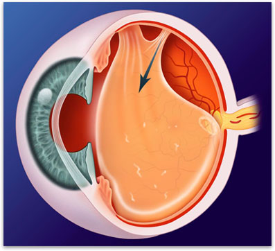
Figure 1. In some people there is an abnormal attachment between the vitreous gel and the retina (arrow), which can result in a torn retina.
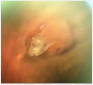
Figure 2. Retinal tear, the vitreous is attached at the lip of the tear.
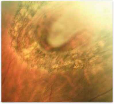
Figure 3. Appearance of a retinal tear after laser treatment.