
Image of the Month - September 2020
(Photo credit: Emily Bishop)
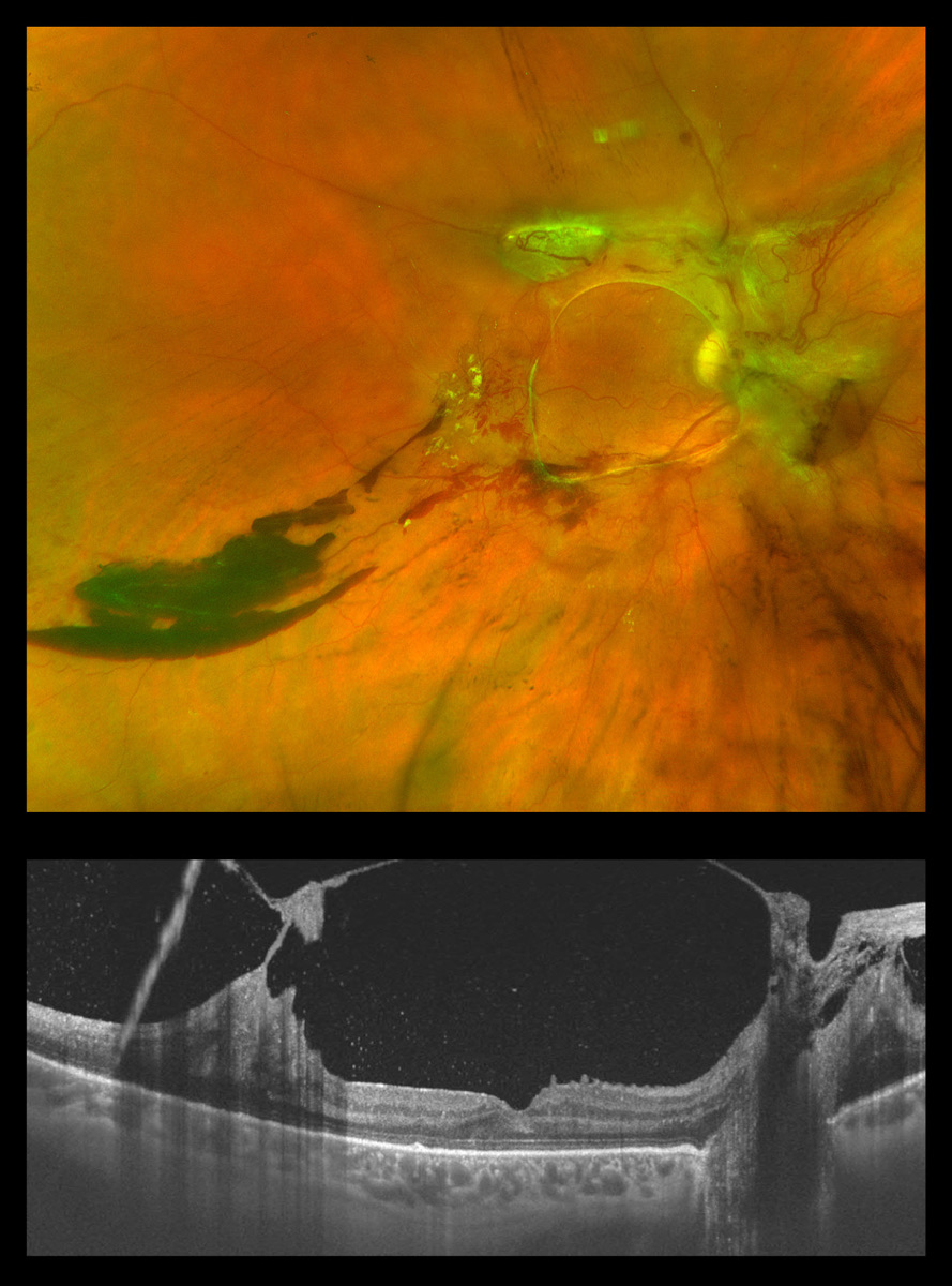
This multi-modal image demonstrates a patient with diabetic tractional retinal detachment with early wolf jaw configuration and vitreous hemorrhage. The fundus photo shows the intense neovascular tractional membranes emanating from the optic disk mostly towards superior and nasal. The cortical hyaloid membranes started to fibrose and contract, giving the wolf jaw appearance. There is some vitreous hemorrhage as well. The non-involved retina looks fairly quiet due to severe ischemia. The OCT shows the membrane attachments to the retinal surface with corresponding traction as well as posterior vitreous red blood cells. Serial anti-VEGF injections with panretinal photocoagulation and staged vitrectomy was recommended.