
Image of the Month - October 2020
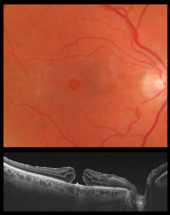
Fundus photography demonstrates a round macular hole with surrounding epiretinal membrane and inner pigment changes. The Optical coherence tomography (OCT) confirms these findings. The hyaloid is absent on the OCT and clinical exam showed a full posterior vitreous detachment.