
Image of the Month - May 2021
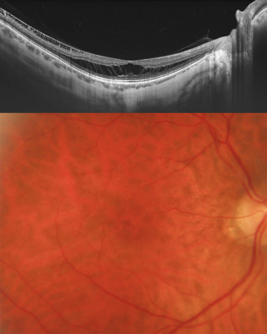
This image series demonstrates a patient with myopic foveoschisis in the right eye. Note the exquisite detail seen in the swept source OCT that demonstrates the cortical vitreous, hyaloid adhesion to the macula, splitting of the retina in multiple layers with residual tissue strands (presumably resembling Mueller cells), a very thin choroid layer which is typical for highly myopic eyes as well as details in the sclera including emissary channels/vessels. The majority of eyes with foveoschisis are being safely observed while a minority undergoes vitrectomy with internal limiting membrane peeling for progressive vision threatening schisis and related symptoms.