
Image of the Month - May 2020
(Photo credit: Jeffrey Barker / Brooke Jocko)
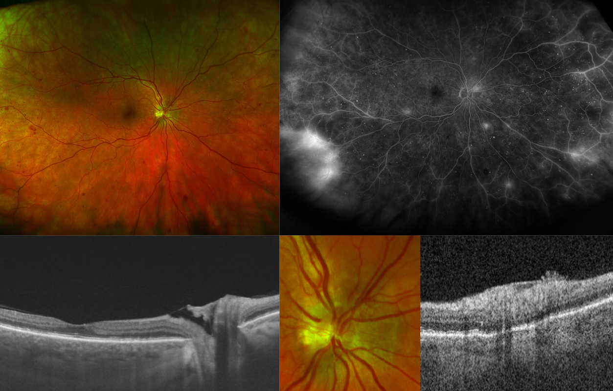
This multimodal image illustrates features of Proliferative diabetic retinopathy. The ultra widefield pseudocolor image demonstrates Neovascularization elsewhere (NVE) and on the disk (NVD) along with hemorrhages in all quadrants (top left). The fluorescein angiography shows corresponding leakage of the neovascularization, as well as significant vascular inflammation and mild capillary non perfusion (top right). The high resolution Swept-source Optical coherence tomography (SS-OCT) line scan demonstrates notably absent macular edema and fibrous vascular tissue within the optic nerve cup (bottom left). The bottom left shows a magnified area of the optic disk with visible NVD and NVE. The NVE is well visible nasally and superior to the disk. The corresponding OCT demonstrates anterior vessel growth towards the vitreous cavity, a hallmark of NV. Ultra widefield fluorescein angiography enhances the examination and has enabled more precise detection and mapping of PDR features. Here, the wide-scan SS-OCT nicely illustrates the corresponding OCT features of NVD and NVE, which can be found overlying the optic nerve cup and growing inwards to the vitreous cavity. This patient was initiated with a hybrid anti-VEGF and panretinal photocoagulation approach and will be followed closely.