
Image of the Month - March 2020
(Photo credit: Stefanie Palmer, C.R.A.)
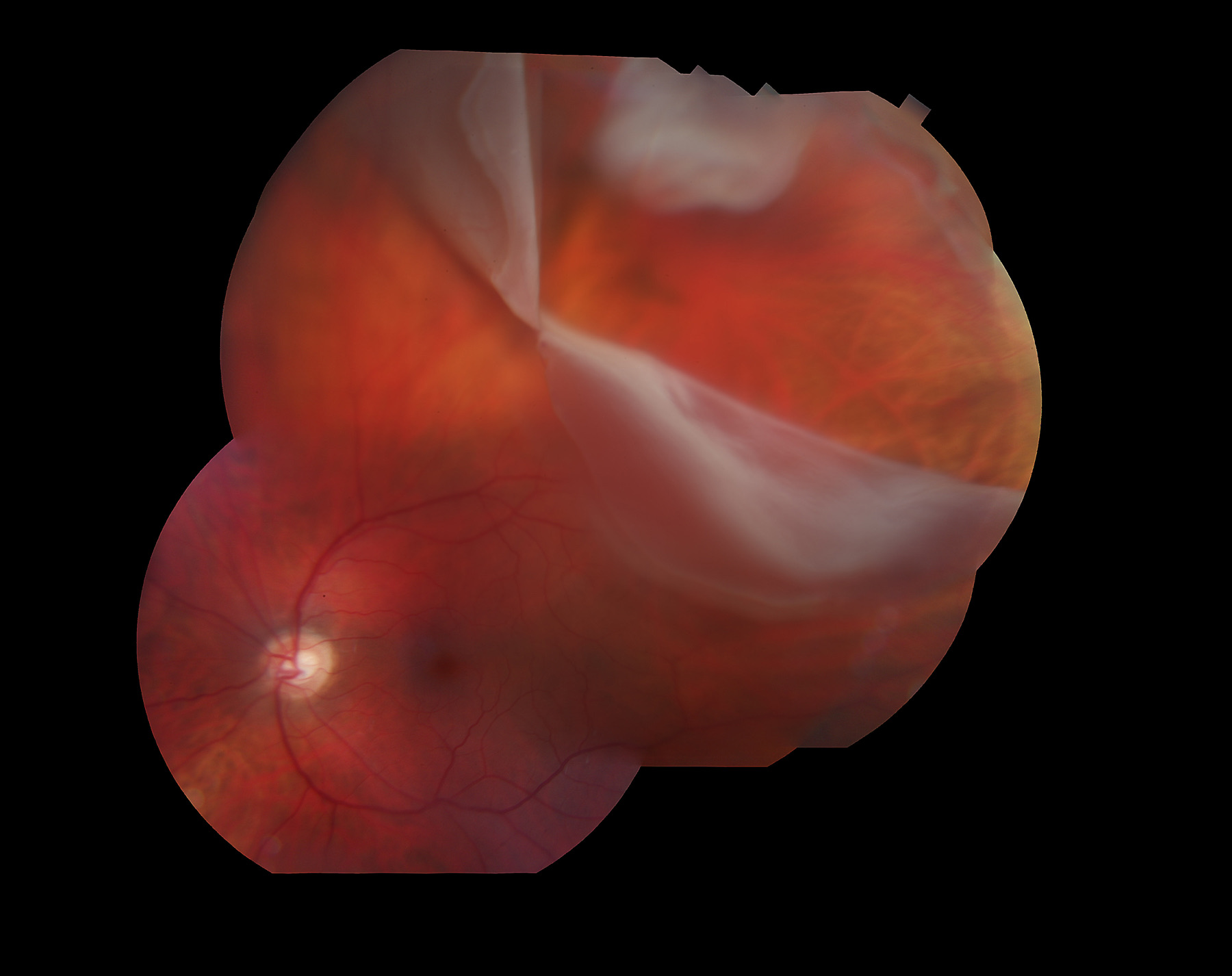
This montage image illustrates a retinal detachment associated with giant retinal tear (GRT). There is a 4 clock hour GRT with partially inverted posterior retinal flap and visible anterior retinal flap in the superotemporal quadrant. Risk factors for GRT include trauma, high myopia and hereditary vitreoretinopathies such as Stickler and Knobloch syndromes as well as others. Management of GRT associated retinal detachment is complex, and usually involves vitrectomy with or without scleral buckle and use of perfluorocarbon liquid (PFCL) to flatten the retina. Close attention to the fellow eye is crucial with careful consideration of prophylactic laser retinopexy in select cases. It is important to distinguish GRT from retinal dialysis. In GRT, the tear occurs in close proximity to the vitreous base with retinal tissue present anteriorly to the tear, whereas in dialysis, the tear occurs at the ora serrata without retinal tissue anteriorly. Retinal dialysis is usually found in younger patients with formed vitreous and has a strong association with blunt trauma. The management is primary scleral buckle in most cases. The patient depicted here has undergone urgent 25-gauge pars plana vitrectomy with diligent vitreous base shaving, PFCL to flatten the retina, fluid gas exchange and 15% C3F8 gas tamponade; and had an excellent anatomical and visual outcome.