
Image of the Month - July 2021
(Photo credit: Stefanie Palmer C.R.A. and Kelsey Cloonan )
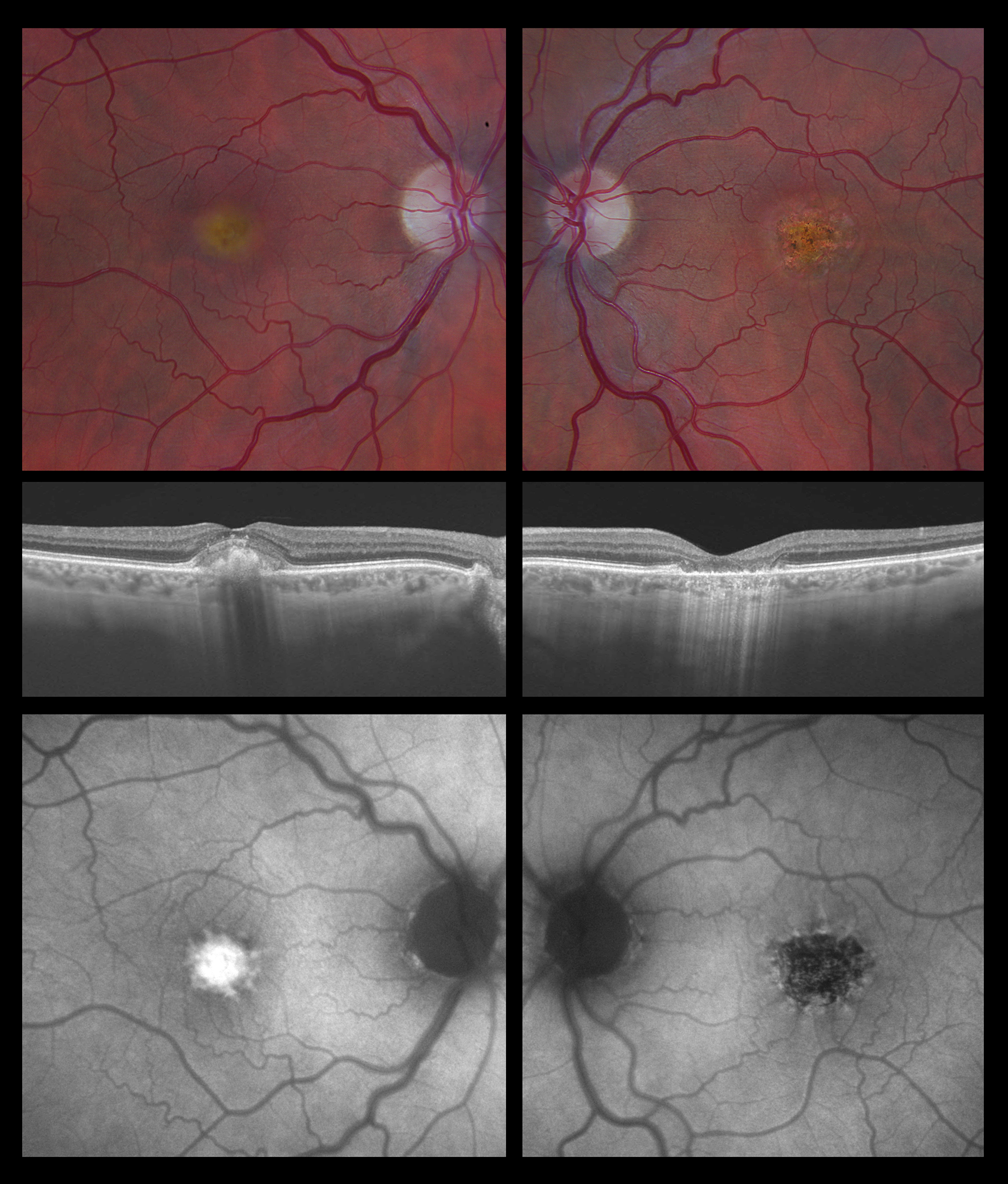
This is an example of two different stages of adult onset vitelliform dystrophy. Classically, vitelliform dystrophy is divided into 5 tages: 1) vitelliform 2) pseudohypoyon 3) vitelliruptive 4) atrophic 5) cicatricial. The right eye shows an elevated subretinal deposit (vitelliform stage). The autofluorescence shows a characteristic hyperautofluorescence spoke-like lesion - the leading hypothesis is that this subretinal material is derived from abnormal accumulation of lipofuscin and hence shows increased autofluorescence. Often times eyes can progress to a breakup of the vitelliform material leading to the pseudohypopyon or vitelliruptive stage (not shown) and this needs to be differentiated from subretinal fluid secondary to exudative macular degeneration. The left eye demonstrates progression to focal geographic atrophy (atrophic stage) with characteristic hypoautofluorescence and loss of outer retinal layers on OCT. Not all eyes with vitelliform dystrophy progress through the different stages and many remain to have fairly good vision in the 20/40 to 20/60 range long-term, but eyes with further progression to atrophy or secondary choroidal neovascularization can fare worse.