
Image of the Month - July 2020
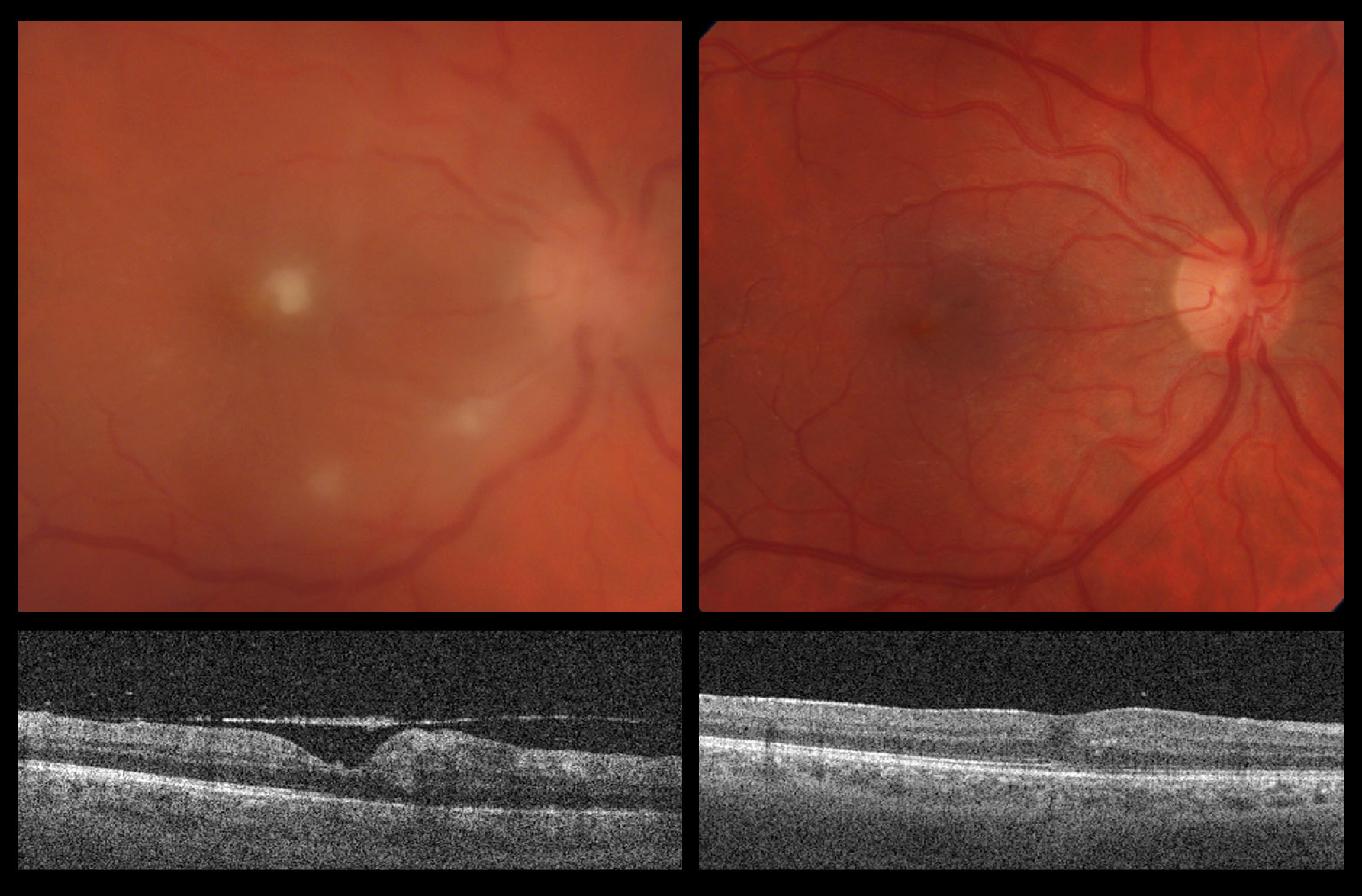
(Photo credit: Jennifer Bahr and Angela Pearson)
The image series here depicts a patient with fungal endophthalmitis. Images on the left depict original presentation with vitritis and fungal lesions, partially in the subhyaloid and subILM space and with connections to the optic disk. Visual acuity was 20/200 and the patient had eye pain for a few days. Upon hospital admission, a systemic workup was conducted along with systemic antifungals and a series of intraviteal voriconazol and amphotericin injections. Despite these efforts, the vitritis progressed. He underwent vitrectomy with induction of posterior vitreous detachment and internal limiting membrane peeling to facilitate removal of all amenable fungal lesions as well as intravitreal injection of amphotericin. Surgical cultures revealed C. albicans. On follow up, the patient continued treatment with systemic antifungals and received a short course of oral steroids. Six week follow up images are depicted on the right side with resolution of vitritis and post surgical macular changes on OCT. Visual acuity improved to 20/40 and the patient was entirely asymptomatic. Fungal endophthalmitis is a vision threatening disease with multiple systemic implications and mainstay of treatment consists of systemic and intravitreal pharmacotherapy. Early surgical intervention is often beneficial and patients need to be worked up for systemic involvement.