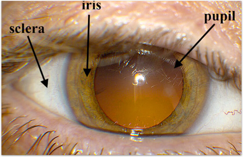
The outer eye
The white part of the eye is the sclera — a tough outer layer that protects the inside of the eye and helps it keep its shape. The colored part is the iris, a circular muscle that controls how much light enters the eye by opening and closing the pupil at its center. The pupil looks dark because of the light-absorbing pigments inside the eye behind it.
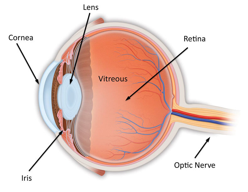
The eye in cross-section
Light enters the eye through the cornea, the clear, curved layer at the very front. It then passes through the pupil and the lens, which focuses the light. Behind the lens, light travels through the vitreous — a clear, jelly-like substance that fills most of the eye — and reaches the retina at the back. The retina converts light into nerve signals, which are sent to the brain through the optic nerve.
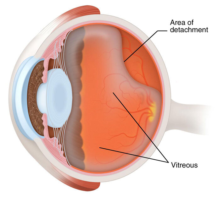
The vitreous
The vitreous is about 99% water, with collagen fibers, salts, sugars, and a small amount of hyaluronic acid that gives it a thicker consistency than water alone. It fills the inside of the eye and helps maintain its shape. It is connected to the retina in only a few places — the macula, the optic nerve, and along the front edge of the retina.
Unlike the fluid in the front part of the eye, the vitreous does not refresh itself over time. As we age, it gradually liquefies and shrinks. This natural change can pull on the retina and is the underlying mechanism behind several conditions retina specialists treat — including retinal tears, retinal detachments, macular holes, and epiretinal membranes.
The retina
The retina is a thin layer of nerve tissue that lines the back of the eye. It is, in fact, an outgrowth of the brain — part of the central nervous system. The retina is often compared to film in a camera, but the analogy understates how complex it is: it has its own blood supply and contains multiple layers of specialized cells that begin processing visual information before sending it to the brain.
The light-sensing cells of the retina come in two types — rods, which handle vision in low light, and cones, which handle color and fine detail.
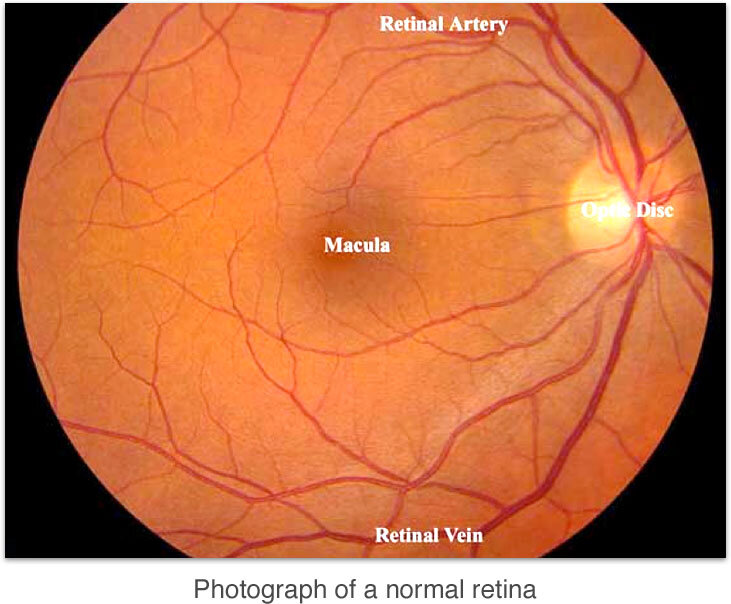
The macula
A small central region of the retina, called the macula, is responsible for the sharp, detailed central vision used for reading, driving, and recognizing faces. The very center of the macula is the fovea, where cones are most concentrated. The fovea contains no rods and no blood vessels — when you look directly at something, you are using your fovea.
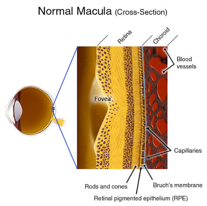
The RPE and the choroid
Beneath the retina sits the retinal pigment epithelium (RPE), a layer of pigmented cells that supports the retina's function. The RPE acts as both an anchor for the retina and a barrier that controls what passes between the retina and the blood vessels behind it.
Behind the RPE is the choroid, a dense bed of blood vessels that supplies oxygen and nutrients to the outer retina. The choroid has one of the highest blood-flow rates of any tissue in the body — more than 70% of the blood in the eye flows through it.