
Image of the Month - January 2020
(Photo credit Jeffrey Barker)
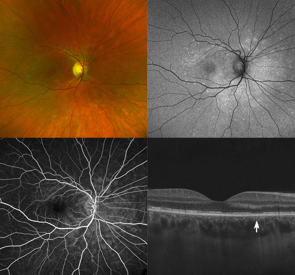
This multi-modal composite image depicts a case of Multiple evanescent white dot syndrome (MEWDS). The pseudocolor widefield photograph (top left) shows multiple, yet subtle small white dots concentrated in the posterior pole of the right eye. Fundus autofluorescence (top right) demonstrates correlating hyper-autofluorescence of the white dots. The Fluorescein angiography image (bottom left) shows characteristic wreath-like staining, and the Optical coherence tomography (OCT; bottom right) image makes evident that the white dots correlate to attenuation of the ellipsoid zone (arrow) on ultra-structural cross sectional imaging. MEWDS is an uncommon, unilateral, inflammatory condition of the retina. It usually affects young to middle aged and otherwise healthy females with myopia. It can be associated with a recent viral prodrome and is self limiting most of the time. Symptoms typically resolve completely a few weeks after onset. The differential diagnosis includes Acute macular neuroretinopathy (AMN), Multifocal choroiditis and panuveitis (MCP), Punctate inner choroidopathy (PIC), Acute zonal occult outer retinopathy (AZOOR) and sarcoidosis. It is important to rule out syphilis, tuberculosis and lymphoma in select cases.