
Fundus Photography
What is Fundus Photography?
Many retinal diseases can change in appearance over time as they progress. It is very important to be able to document any changes in order to tell if treatment is needed. A good way to document the appearance of the retina is to use fundus photography.
All of our offices have the capability of obtaining high-resolution images of the retina. We use fundus photography to document the appearance of retinal abnormalities identified during the examination. The high quality images are captured digitally and stored at RVS to be used as a point of reference for future comparison.
The digital images we obtain can be further refined and analyzed using computer software to focus on specific areas of interest, plan for laser treatment or generate montages.
Fundus photography is also often used in conjunction with Fluorescein angiography to aid in the diagnosis and treatment of many medical retinal disorders such as diabetic retinopathy, age related macular degeneration, other retinal circulatory disturbances or ocular tumors.
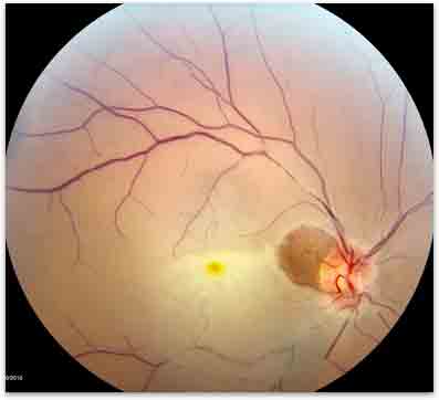
Figure 1. Central retinal artery occlusion.
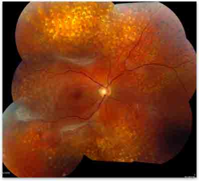
Figure 2. Severe diabetic retinopathy.